Liposome Structure Diagram: How to Draw It Correctly
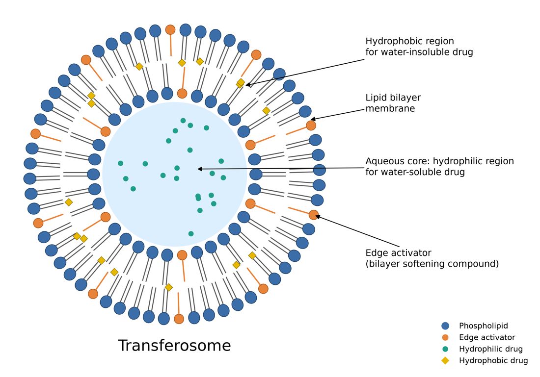
A liposome diagram looks simple, but it is one of the most common places where pharmacy projects and theses lose marks: heads facing the wrong way, drugs in the wrong compartment, or a "liposome" that is really a micelle. This guide shows what a correct diagram must include.
1. The basic structure
- Phospholipid bilayer: two layers of phospholipids. The polar heads face water on both sides (the outside medium and the inner core). The non-polar tails point into the middle of the bilayer, towards each other.
- Aqueous core: the water-filled space in the centre.
- Cholesterol (often shown as small rings between the tails): makes the bilayer more rigid and less leaky.
A micelle is different: it has only one layer, tails in the centre, and no aqueous core. Do not draw one when you mean a liposome.
2. Where the drugs go
- Hydrophilic (water-soluble) drugs sit in the aqueous core, for example doxorubicin in Doxil.
- Lipophilic (fat-soluble) drugs sit inside the bilayer, between the tails, for example amphotericin B in AmBisome.
- Targeting ligands and PEG (in stealth liposomes) are attached to the outer surface.
Use two different colours for the two drug types and add a small legend.
3. Types of liposomes by size and lamellarity
- SUV (small unilamellar vesicle): one bilayer, about 20 to 100 nm.
- LUV (large unilamellar vesicle): one bilayer, larger than 100 nm.
- MLV (multilamellar vesicle): several bilayers like onion layers, about 0.5 to 5 µm.
4. Related vesicles (and how to show the difference)
- Niosome: same bilayer shape, but made of non-ionic surfactants (such as Span 60) with cholesterol instead of phospholipids.
- Transfersome: phospholipids plus an edge activator (such as sodium cholate or Tween 80), which makes the vesicle ultra-deformable so it can squeeze through skin pores.
- Ethosome: phospholipids with a high amount of ethanol (about 20 to 45%), which makes the vesicle soft and improves skin penetration.
Checklist before you submit
- Heads face water on both sides; tails face each other.
- Hydrophilic drug in the core, lipophilic drug in the bilayer.
- Labels spelled correctly (phospholipid, cholesterol, aqueous core).
- Exported at 300 dpi or as vector (PDF/SVG).
Make it in one minute
Type "liposome structure with hydrophilic drug in the core and lipophilic drug in the bilayer" into SciFigures and get a clean, correctly labelled diagram. The free plan gives 3 figures a month. Try the drug delivery diagram maker or the nanoparticle diagram maker.
See also figures for a pharmacy project or thesis and drug release kinetics models.
More guides
- How to Make a Graphical Abstract (Step by Step)
- Journal Figure Requirements: Size, DPI and File Formats
- How to Make Good Scientific Figures for Your Thesis
- Colour-Blind Friendly Colour Palettes for Scientific Figures
- How to Draw a Mechanism of Action Diagram
- How to Edit SVG Figures in PowerPoint, Illustrator and Inkscape
- Free BioRender Alternatives for Scientific Figures
- How to Fix AI-Generated Scientific Images Before Using Them
- How to Make a Volcano Plot (and Read One)
- How to Calculate IC50 from a Dose-Response Curve
- Kaplan-Meier Survival Curve Explained (with Example)
- Drug Release Kinetics Models: Zero Order, First Order, Higuchi, Korsmeyer-Peppas
- How to Make a Forest Plot for a Meta-Analysis
- SD vs SEM Error Bars: Which Should You Use?
- How to Draw a Research Methodology Flowchart
- Figures for a B.Pharm or M.Pharm Project and Thesis
- Free Scientific Figure Maker for a Thesis
- How to Make 300 DPI Figures for a Journal
- Best Tools for Research Paper Figures (2026)
- BioRender vs SciFigures: Which Should You Use?
- Best Free Graphical Abstract Makers (2026)